Measurement of Dye Concentration
In microscopy, the purpose of image processing is not just the generation of good-looking images but also the derivation and extraction of quantitative or at least qualitative results. The algorithm of color deconvolution serves this purpose. It converts color images of stained tissue samples into concentration distributions of dye uptake.
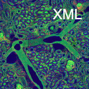
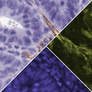
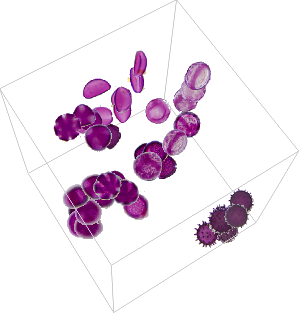
The tissue in the following image has been stained using Hematoxylin C19 and DAB (3,3'-Diaminobenzidine).
Literature provides the typical RGB colors of the two stains. Theoretically, the dye colors are obtained by multiplying their color spectrum with the sensitivity spectrum of the red, green and blue channels of the CCD in the recording camera. Calling these multiplications a color convolution results in the inverse operation being called a color deconvolution, which is effectively a color demixing of stain colors.
Obtain the transformation matrix from staining dye to RGB color channels.
Compute the linear inverse transformation from color to dye concentration.
The actual demixing is performed in the log-scale of color intensities, since the color absorption is exponentially proportional to the dye concentration. A short implementation of color deconvolution is given here.
Perform color deconvolution into Hematoxylin and DAB dye concentration.
Multiply each component with the color of its corresponding stain.
Representing the dye concentration with false colors.