The Wolfram Solution forMedical ImagingUse built-in functions for segmentation, registration, restoration, and analysis of 2D and 3D volumetric images; prototype new algorithms quickly and efficiently; and deploy tools as standalone or web-based applications—from one system. The Wolfram medical imaging solution provides a complete integrated workflow for image processing and application development, with the speed and performance benefits of GPU computation, parallel processing, and out-of-core technology. |
 |
The Wolfram Edge
How Wolfram Compares
Key Capabilities
Wolfram technologies include thousands of built-in functions and curated data on many topics that let you:
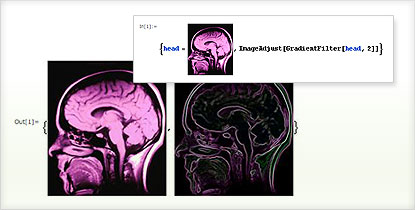
- Design software programs to do edge-preserving smoothing, denoising, sharpening, and other enhancements
- Visualize tomography data in 2D or 3D such as CT and MRI scans
- Slice through 3D data and explore the inside of a volume
- Create pattern-recognition algorithms for computer-aided diagnosis or tumor detection
- Develop and simulate radio-frequency pulse sequences
- Compare imaging measurements with biological models
- Scan cell samples for abnormalities
- Study videos of runners to improve the efficiency of their movements
- Deblur CT slices and remove inhomogeneities from MRI backgrounds
- Capture and process images from imaging devices in real time
- Analyze fiber orientation in lab-grown tissue to determine its strength
- Use noninvasive techniques to study the heart, reducing risk to the patient
- Deploy web applications for remote diagnosis

Using blob-analysis techniques to find cells in a microscopic image

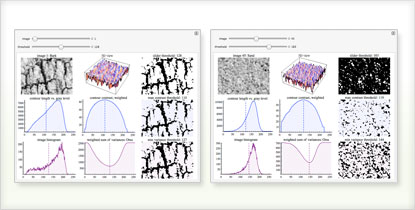
Creating interactive tools for analyzing images


